

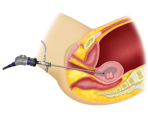
Diagnostic hysteroscopy is a minimally invasive procedure used to examine the inside of the uterus (uterine cavity) using a hysteroscope – a thin, telescope-like instrument with a camera and light source. The hysteroscope is passed through the vagina and cervix, without any abdominal incisions, allowing the doctor to directly visualise the uterine lining and identify any abnormalities.
The suitability of diagnostic hysteroscopy depends on several individual factors, including the reason for evaluation, menstrual and fertility history, previous uterine or cervical procedures, and overall reproductive health.
When is Diagnostic Hysteroscopy Considered?
Diagnostic hysteroscopy may be considered for women with:
- Abnormal or heavy uterine bleeding.
- Irregular menstrual cycles.
- Postmenopausal bleeding.
- Suspected uterine polyps, fibroids, or adhesions.
- Recurrent pregnancy loss or unexplained infertility.
- Suspected congenital abnormalities of the uterus.
- Abnormal findings on ultrasound or other imaging that require direct visual assessment.
- Follow-up evaluation prior to fertility treatments, such as IVF.
Before recommending the procedure, the doctor may evaluate:
- The patient’s menstrual history and presenting symptoms.
- Findings from prior ultrasound or imaging studies.
- Fertility history and reproductive goals, where relevant.
- Previous uterine, cervical, or pelvic surgical history.
- Overall health and any factors affecting suitability for the procedure.
A detailed evaluation helps determine whether diagnostic hysteroscopy is appropriate and what findings it may help clarify.
What Happens During Diagnostic Hysteroscopy?
Before the procedure, the doctor reviews the patient’s medical history and the reason for evaluation, along with any prior imaging. The procedure may be performed under local anaesthesia, sedation, or general anaesthesia, depending on individual circumstances and whether it is combined with other procedures.
During the procedure, the hysteroscope is gently passed through the vagina and cervix into the uterine cavity, without any incisions. A sterile fluid or gas is used to gently distend the uterine cavity, allowing for a clear view of the uterine lining, the openings of the fallopian tubes, and any abnormalities such as polyps, fibroids, adhesions, or structural irregularities. If a biopsy is required, a small tissue sample may be taken for further evaluation.
The exact approach and any additional steps depend on the findings and the reason for the procedure.
Recovery After Diagnostic Hysteroscopy
Recovery after diagnostic hysteroscopy is generally quick, as it does not involve any incisions. Patients are generally advised to follow their doctor’s postoperative instructions carefully.
Important aspects of recovery may include:
- Expecting mild cramping or light spotting for a short period after the procedure.
- Taking prescribed or over-the-counter medications for mild discomfort, if needed.
- Resuming most daily activities within a short time, as advised by the doctor.
- Avoiding sexual intercourse or use of tampons for a short period, if advised.
- Attending follow-up appointments to discuss findings and any further treatment needed.
Your doctor will provide personalised guidance based on the procedure performed and your overall health.
FAQs
1. Is diagnostic hysteroscopy painful?
Most women experience only mild discomfort or cramping during the procedure, similar to menstrual cramps. The level of discomfort can vary depending on whether it is performed with local anaesthesia, sedation, or general anaesthesia
2. How long does a diagnostic hysteroscopy take?
The procedure itself is generally quick, often taking a short time to complete, though this can vary depending on the findings and whether a biopsy or additional evaluation is needed.
3. Can diagnostic hysteroscopy be done in an outpatient setting?
Yes, in many cases, diagnostic hysteroscopy can be performed as an outpatient procedure, allowing the patient to return home the same day. Your doctor will advise on the most suitable setting based on your individual case.
4. Will I need anaesthesia for diagnostic hysteroscopy?
This depends on individual factors and clinical preference. Some procedures are performed with little to no anaesthesia, while others may require local anaesthesia, sedation, or general anaesthesia, particularly if combined with other procedures.
5. What happens if an abnormality is found during diagnostic hysteroscopy?
If a treatable abnormality, such as a polyp or fibroid, is identified, your doctor may proceed with operative hysteroscopy to address it during the same procedure or recommend a follow-up procedure, depending on the findings and your individual situation.