

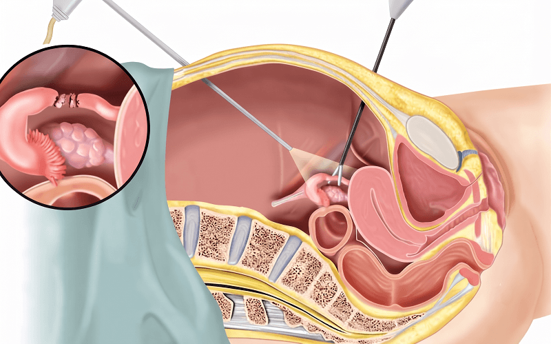
Diagnostic hysteroscopy is a minimally invasive procedure used to examine the inside of the uterus (womb) using a thin, lighted instrument called a hysteroscope, which is passed through the vagina and cervix. It helps identify the cause of issues like abnormal uterine bleeding, infertility, recurrent miscarriage, or suspected polyps and fibroids — without any abdominal incision.
How to Prepare for Diagnostic Hysteroscopy
The procedure is usually scheduled shortly after your menstrual period ends, when the uterine lining is thinnest and easiest to view. Your doctor may recommend a pregnancy test beforehand, and you should inform them of any allergies, medications, or existing health conditions. If sedation or anesthesia is planned, you may be asked to fast for a few hours and arrange for someone to accompany you home afterward.
What Happens During Diagnostic Hysteroscopy
After positioning and inserting a speculum, the doctor gently passes the hysteroscope through the cervix into the uterus. A sterile fluid or gas is used to gently expand the uterine cavity, allowing a clear view on a connected monitor. The doctor examines the uterine lining for abnormalities such as polyps, fibroids, scar tissue, or a septum, and may take a small tissue sample (biopsy) if needed. The procedure typically takes 15–30 minutes and, depending on the case, can often be done under local anesthesia or mild sedation as a day-care procedure.
Benefits of Diagnostic Hysteroscopy
- Minimally invasive, with no abdominal incision required.
- Quick recovery, often allowing same-day discharge.
- Direct visualization of the uterine cavity for more accurate diagnosis than ultrasound alone.
- Can sometimes combine diagnosis with minor treatment in the same sitting.
- Low risk of complications when performed by a trained specialist.
However, suitability depends on individual health factors, and your doctor will confirm if it is the right diagnostic approach for you.
Diagnostic vs Operative Hysteroscopy
Diagnostic hysteroscopy is primarily used to view and identify problems inside the uterus. If an abnormality such as a polyp, fibroid, or adhesion is found and needs removal or correction, your doctor may recommend proceeding to (or scheduling separately) an operative hysteroscopy, which treats the condition using specialized instruments. The right approach depends on findings during the examination and the patient’s overall clinical picture.
FAQs
1. Is diagnostic hysteroscopy painful?
Most women experience only mild cramping, similar to menstrual cramps, during the procedure. Local anesthesia or mild sedation is often used to minimize discomfort, and the procedure is generally well-tolerated.
2. How long does it take to recover after the procedure?
Recovery is usually quick. Most women can resume normal activities within a day, though some light spotting or mild cramping may continue for a day or two. Since it’s often a day-care procedure, you can typically go home the same day.
3. Is anesthesia required for diagnostic hysteroscopy?
Not always. Many diagnostic hysteroscopies can be performed with little to no anesthesia, using just local anesthesia or mild sedation. The choice depends on your doctor’s assessment, your comfort level, and whether any additional procedures (like a biopsy) are planned.
4. What are the risks associated with diagnostic hysteroscopy?
It is a low-risk procedure overall, but rare complications can include infection, bleeding, or, very rarely, injury to the uterus or cervix. Your doctor will discuss your specific risk factors before the procedure.
5. When can I expect the results, and what happens next?
If only a visual examination is done, your doctor may share initial findings right after the procedure. If a biopsy was taken, results typically take a few days to a couple of weeks, depending on the lab. Based on the findings, your doctor will discuss whether further treatment, such as operative hysteroscopy, is needed.